In a move that may eliminate the need for painful biopsies for people with kidney disease, London-based researchers have found a way to accurately gauge the amount of salt within the organ.
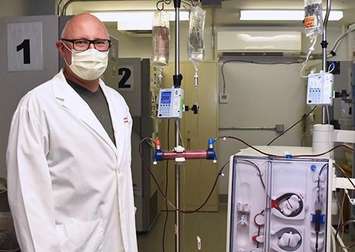
Scientists at Lawson Health Research Institute developed new technology and software in order to adapt a PET/MRI machine at St. Joseph’s Health Care London to complete the difficult imaging task.
“Salt within the kidneys have only been imaged in pre-clinical models, and low weight, healthy volunteers,” said Dr. Christopher McIntyre, Lawson Scientist and Nephrologist at London Health Sciences Centre. “Since the kidney is further away from the MRI coils, and the organ moves when a person breathes, it is definitely very hard to image.”
In the first of its kind study, researchers using the new imagining technology examined salt within the kidneys of ten healthy volunteers with different body types, five patients with kidney disease and patients who had a combination of kidney disease and heart failure. For treatment, it is important for individuals with kidney disease specifically to be able to release salt and water.
Currently, the only way for physicians to get an idea of salt levels in a patient is to perform a kidney biopsy.
"The problem is that the biopsies are painful, they have risks, and because it is a small sample of the kidney, we don’t get an accurate perspective of the kidney as a whole,” said McIntyre. “This will now allow us to diagnose and manage both chronic and acute kidney disease. It is a significant step forward.”
The study findings have been published in the journal Radiology.
In the next part of their research, the Lawson team will compare salt MRI findings to biopsies and examine potential new therapies.
“We are hoping we will have a higher degree of certainty moving forward to predict what will happen within the kidneys of these patients, with the possibility of using new targeted and effective treatments in the future,” said McIntyre.